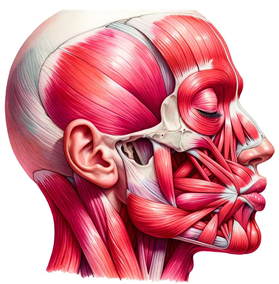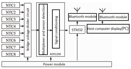

This paper delves deeply into the innovative realm of integrating human emotions with wearable technology. The primary focus is on the conceptualization and development of a kiss transfer device that harnesses the power of wearable technology to bridge the physical gap in human-human interactions. By investigating the intricate nuances of the human-human kissing process, the research seeks to replicate this intimate gesture through a technological medium. The paper not only elaborates on the anatomy, evolution, and hormonal dynamics of kissing but also underscores the transformative potential of wearable technology in capturing and transmitting these intimate moments. This exploration opens up new horizons for long-distance relationships, offering a tangible touchpoint that goes beyond traditional communication methods. Through this pioneering work, the research positions wearable technology as not just a tool for communication but as an extension of our human emotions and expressions.

Simulation study of calcaneal insertion in the treatment of children’s flat foot
Vol 1, Issue 2, 2020
Download PDF
Abstract
One of the correction methods of children’s flat foot is calcaneal insertion. The purpose of this work is to determine the performance of calcaneal stop by using two bioco MPatible materials. The Finite Element Method (FEM) is used. The material analysis is AISI 316L steel and Ti-6Al-4V titanium alloy. Using Cuban anthropometric models, loads were calculated based on foot biomechanics and body weight of boys and girls aged 10 and 12. The results show that the implant model ensures the mechanical strength of the two materials. The difference between the two stresses is 4.44 MPa, accounting for 5% of the difference. Both materials have sufficient mechanical strength reserves because the maximum stress is less than the elastic limit of the material and ensures the mechanical strength of the calcaneal stop design.
Keywords
References
- Pavone V, Vescio A, Di Silvestri CA, et al. Outcomes of the calcaneo-stop procedure for the treatment of juvenile flatfoot in young athletes. Journal of Pediatric Orthopedics 2018; 12(6): 582–589.
- Calvo S, Marti CR, Rasero PM, et al. Más de 10 años de seguimiento de la técnica de calcáneo stop [Calcaneal insertion technique was followed up for more than 10 years]. Revista Española de Cirugía Ortopédica y Traumatología 2016; 60(1): 75–80.
- Pinho CF, Costa G, Santos CM, et al. Long term outcomes of calcaneo-stop procedure in the treatment of flexible flatfoot in children: A retrospective study. Acta Med Port 2017; 20(7–8): 541–545.
- Samaila E, Ggelmini M, Invernizzi E, et al. Low incidence of complications of arthroereisis with Calcaneo-Stop at long term follow-up. Foot and Ankle Surgery 2017; 23(Suppl.1): 71.
- Fleites Lafont LM, Oscar Marrero Riverón CL, Alcalá Alfonzo EJ. Técnica calcáneo-stop con elongación de tendones peroneos en el pie plano de pacientes con parálisis cerebral infantil. [Calcaneal-stop technique with peroneal tendon elongation in flat feet in patients with infantile cerebral palsy]. Revista Cubana de Ortopedia y Traumatología 2014; 28(1): 39 57.
- Giannini S, Cadossi M, Mazzotti A, et al. Bioabsorbable calcaneo-stop implant for the treatment of flexible flatfoot: a retrospective cohort study at a minimum follow-up of 4 years. Journal of Foot and Ankle Surgery 2017; 56(4): 776 782.
- Faldini C, Mazzotti A, Panchera A, etc. Patient-perceived outcomes after subtalar arthroereisis with bioabsorbable implants for flexible flatfoot in growing age: A 4-year follow-up study. European Journal of orthopedic surgery and Traumatology 2018; 8(4): 707–712.
- Calderín Pérez B, González Carbonell RA, Landín Sorí M, et al. Aplicabilidad de la simulación computacional en la biomecánica del disco óptico [Application of computational simulation in optic disc biomechanics]. Arch Med Camagüey 2015; 19(1): 73–82.
- Calderín Pérez B, González Carbonell RA, Landín Sorí M, et al. Análisis biomecánico del disco óptico bajo la variación de presión intraocular y rigidez escleral [Biomechanical analysis of optic disc under changes of intraocular pressure and scleral hardness]. Rev. Cubana. Inv. Bioméd 2016; 35(2): 136–157.
- Pérez Pozo L, Briones Picheira F, Aguilar Ramírez C. Análisis de esfuerzos mediante el método de elementos finitos de implantes dentales de titanio poroso [The stress of porous titanium implant was analyzed by finite element method]. Ingeniería y Desarrollo 2015; 33(1): 80–97.
- Cárdenas Oliveros JA, Cárdenas Caña JH, Teixeira Da Silva JM. Tornillo intrapedicular y prisionero. Análisis por el Método de Elementos Finitos [Intrapedicular and stud bolt. Analysis by the finite element method]. Ingeniería Mecánica 2017; 20(3): 129–135.
- Ouyang H, Deng Y, Xie P, et al. Biomechanical comparison of conventional and optimised locking plates for the fixation of intraarticular calcaneal fractures: a finite element analysis. Comput Methods Biomech Biomed Engin 2017; 20(12): 1339–1349.
- Wong D, Niu W, Wang Y, et al. Finite element analysis of foot and ankle impact injury: Risk evaluation of calcaneus and talus fracture. PLoS ONE 2016; 11(4): 1–14.
- Shih Cherng L, Carl Pai-Chu C, Simon Fuk-Tan T, et al. Stress distribution within the plantar aponeurosis during walking—A dynamic finite element analysis. Journal of Mechanics in Medicine & Biology 2014; 14(4): 1–17.
- Telfer S, Erdemir A, Woodburn J, et al. What has finite element analysis taught us about diabetic foot disease and its management? A systematic review. PLoS ONE 2014; 9(10): e109994.
- Guiotto A, Sawacha Z, Guarneri G, et al. 3D finite element model of diabetic neuropathy foot: A Gait analysis driven approach. Journal of Biomechanics 2014; 47(12): 3064–3071.
- González Carbonell RA, Álvarez García E, Campos Pérez Y. Tacón de torque. Análisis tensional y deformacional utilizando el Método de Elementos Finitos [Torque heel. Stress and deformation analysis using the Finite Element Method]. Ingeniería Mecánica 2007; 10(2): 79–83.
- Roth S (inventor). Titanium implants injected to correct children’s flat feet. USA patent: US 8, 267,977 B2. 2012.
- Álvarez Sintes R. Medicina General Integral [Comprehensive General Medicine]. 3rd ed. Havana: Medical Science Press, 2014.
- Álvarez C, Palma VW. Desarrollo y biomecánica del arco plantar [Development and biomechanics of plantar arch]. Ortho-tips 2010; 6(4): 215–222.
- Viladot Voegeli A. Anatomía funcional y biomecánica del tobillo y el pie [Functional anatomy and biomechanics of ankle and foot]. Rev Esp Reumatol 2003; 30(9): 469–477.
- Elias CN, Lima JHC, Valiev R, et al. Biomedical applications of titanium and its alloys. JOM 2008; 60(3): 46–49.
- Disegi J. Implant materials Wrapped with 18% chromium, 14% nickel and 2.5% molybdenum stainless steel. Synthes, West Chester, USA, 2009. Accessed: Jan 20, 2018. [Online]. Available from: https://docplayer.net/22402352-Third-edition-implant-materials-wrought-18-chromium-14-nickel-2-5-molybdenum-stainless- steel.html.
- Bartolomeu F, Buciumeanu M, Pinto E, et al. 316L stainless steel mechanical and tribological behavior—A comparison between selective laser melting, hot pressing and conventional casting. Additive Manufacturing 2017; 16: 81–89.
- Sierra Uribe JH, Bravo Molina, OM, Acevedo Peña P, et al. Evaluación electroquímica de recubrimientos de biovidrio/Al2O3 soportados sobre acero inoxidable AISI 316L y su relación con el carácter bioactivo de las películas [Electrochemical Evaluation of Bioglass / Al2O3 Coating on AISI 316L Stainless Steel and Its Relationship with Bioactivity of Films]. Revista Latinoamericana de Metalurgia y Materiales 2015; 35(2): 151–164.
Supporting Agencies
Copyright (c) 2020 Elsa Nápoles-Padrón, Juan Pablo Pacheco-González, Raide A. González-Carbonell, Armando Ortiz-Prado, Jesús Hernández-de la Torre

This work is licensed under a Creative Commons Attribution 4.0 International License.

Prof. Zhen Cao
College of Information Science & Electronic Engineering, Zhejiang University
China, China
Processing Speed
-
-
-
- <5 days from submission to initial review decision;
- 62% acceptance rate
-
-
Asia Pacific Academy of Science Pte. Ltd. (APACSCI) specializes in international journal publishing. APACSCI adopts the open access publishing model and provides an important communication bridge for academic groups whose interest fields include engineering, technology, medicine, computer, mathematics, agriculture and forestry, and environment.


